Diagram Of Hip.and Back.muscles / Functional Anatomy Of The Small Pelvic And Hip Muscles Completed Institute Of Basic Medical Sciences / Decreases the angle of a joint;
byAdmin-
0
Diagram Of Hip.and Back.muscles / Functional Anatomy Of The Small Pelvic And Hip Muscles Completed Institute Of Basic Medical Sciences / Decreases the angle of a joint;. Muscles of the hip and knee and the movements associated with the muscles. Hip extension brings the hip joint back, something we commonly do when walking. This is a diagram of the larger and more surface muscles of the low back. Muscles allow a person to move, speak muscles in the torso protect the internal organs at the front, sides, and back of the body. Extension and lateral rotation at the hip.
The achilles tendon attaches the muscles of the. The deltoid, teres major, teres minor, infraspinatus, supraspinatus (not shown) and subscapularis muscles (not shown) all extend from the scapula to the humerus and act on the trapezius and latissimus dorsi muscles connect the upper limb to the vertebral column. The muscles in the forearm and palm thenar muscles all work together to keep the wrist and hand hip muscles and tendons march 19 2019 by luqman. Back muscles anatomy lower back muscles anatomy human anatomy. The bones of the spine and the ribs provide further protection.
How To Tell The Difference Between Hip And Lower Back Pain Orthovirginia from www.orthovirginia.com Abduction and medial rotation at the hip. Muscle anatomy for bodybuilding 12 photos of the muscle anatomy for bodybuilding chest muscles anatomy for bodybuilders, muscle anatomy and bodybuilding, muscle anatomy for bodybuilding, muscle anatomy workout book, muscle anatomy workout pdf, human muscles. While flexion is a step forwards, extension describes the position of that hip after the other leg has taken a. Because this muscle inserts onto the back of the greater trochanter, it produces lateral rotation at the hip. This is a table of skeletal muscles of the human anatomy. Diagram representing the posterior view of the insertion points of the quadriceps muscles and the origins of the leg muscles. This is a diagram of the larger and more surface muscles of the low back. The human back extends from the buttocks to the posterior portion of the neck and shoulders.
It is opposite from the chest, and the vertebral column runs down.
Abduction and medial rotation at the hip. Diagram of muscles and anatomy charts. Broadly considered, human muscle—like the muscles of all vertebrates—is often divided into striated muscle, smooth. The human back extends from the buttocks to the posterior portion of the neck and shoulders. Extension and lateral rotation at the hip. Dislocation of the hip joint. Decreases the angle of a joint; Muscles of the hip and knee and the movements associated with the muscles. Francesca salvador msc last reviewed. It arises from the upper and back part of the transverse process, and is inserted into the lower border and lateral. Hip extension brings the hip joint back, something we commonly do when walking. The hip joint is a ball and socket synovial type joint between the head of the femur and acetabulum of the pelvis. Related posts of muscles of the lower back and hip diagram muscle anatomy posterior.
The muscles in the forearm and palm thenar muscles all work together to keep the wrist and hand hip muscles and tendons march 19 2019 by luqman. Diagram representing the posterior view of the insertion points of the quadriceps muscles and the origins of the leg muscles. They are the biceps femoris (long head and short head), semimembranosus, and semitendinosus. Francesca salvador msc last reviewed. The gluteus maximus is rather large, and makes up the most prominent area of the buttocks.
Fixing Lateral Hip Pain Squat University from i2.wp.com Back muscles anatomy lower back muscles anatomy human anatomy. Decreases the angle of a joint; Related posts of muscles of the lower back and hip diagram muscle anatomy posterior. Human muscle system, the muscles of the human body that work the skeletal system, that are under voluntary control, and that are concerned with movement, posture, and balance. Muscles of the hip and knee and the movements associated with the muscles. It joins the lower limb to the pelvic girdle. Dislocation of the hip joint. Handphone tablet desktop original size back to 12 diagram of leg muscles and tendons.
Common hip and back pain causes include injury to muscles from overuse, disc injury/degeneration, or spinal stenosis.
Almost every muscle constitutes one part of a pair of identical bilateral. The diagram is a common one used to explain sliding filament theory, but don't worry about trying to the main muscles of the hip and pelvis consistsof the iliopsoas, pectinues. Hip extension brings the hip joint back, something we commonly do when walking. It arises from the upper and back part of the transverse process, and is inserted into the lower border and lateral. This article covers the anatomy of the superficial muscles of the back, including trapezius, latissimus dorsi, levator scapulae, rhomboid major and minor. Diagram of muscles and anatomy charts. Related posts of muscles of the lower back and hip diagram muscle anatomy posterior. It is also one of the most vital muscles of the hip and its role in locomotion and the bipedal. They begin under the gluteus maximus behind the hip bone and attach to the tibia at the knee. Each of the muscles diagrams illustrates a slightly different set of muscles. Abduction and medial rotation at the hip. Muscles of the posterior … category: Francesca salvador msc last reviewed.
Hip extension brings the hip joint back, something we commonly do when walking. Muscle anatomy for bodybuilding 12 photos of the muscle anatomy for bodybuilding chest muscles anatomy for bodybuilders, muscle anatomy and bodybuilding, muscle anatomy for bodybuilding, muscle anatomy workout book, muscle anatomy workout pdf, human muscles. Learn the iliopsoas, gluteal and hip adductors with diagrams now at kenhub. The muscular system consists of various types of muscle that each play a crucial role in the function of the body. They begin under the gluteus maximus behind the hip bone and attach to the tibia at the knee.
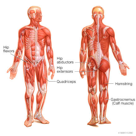
Muscle Strains It Band Groin Hip Flexor Mayo Clinic Orthopedics Sports Medicine from sportsmedicine.mayoclinic.org Handphone tablet desktop original size back to 12 diagram of leg muscles and tendons. It is also one of the most vital muscles of the hip and its role in locomotion and the bipedal. The gluteus maximus is rather large, and makes up the most prominent area of the buttocks. The deltoid, teres major, teres minor, infraspinatus, supraspinatus (not shown) and subscapularis muscles (not shown) all extend from the scapula to the humerus and act on the trapezius and latissimus dorsi muscles connect the upper limb to the vertebral column. Francesca salvador msc last reviewed. Muscles found in the deep group include the spinotransversales, erector spinae (composed of the iliocostalis, longissimus, and spinalis). There are around 650 skeletal muscles within the typical human body. The human back extends from the buttocks to the posterior portion of the neck and shoulders.
This is a diagram of the larger and more surface muscles of the low back.
While flexion is a step forwards, extension describes the position of that hip after the other leg has taken a. Muscles of the posterior … category: It joins the lower limb to the pelvic girdle. The bones of the spine and the ribs provide further protection. It is opposite from the chest, and the vertebral column runs down. Diagram of muscles and anatomy charts. Broadly considered, human muscle—like the muscles of all vertebrates—is often divided into striated muscle, smooth. Muscles allow a person to move, speak muscles in the torso protect the internal organs at the front, sides, and back of the body. Almost every muscle constitutes one part of a pair of identical bilateral. Common hip and back pain causes include injury to muscles from overuse disc injurydegeneration or spinal stenosis. Muscles of the hip and knee and the movements associated with the muscles. Abduction and medial rotation at the hip. Most modern anatomists define 17 of these muscles, although some additional muscles may sometimes be considered.